Structure of a hantavirus protein as a promising model for drug design
Posted by Acubiz | BlogX-ray crystallography provides drug template against disease transmitted by small rodents
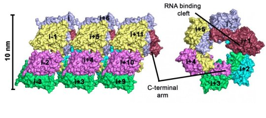
Left: Hexameric rings form a tube of viral capsid. Right: view from the other side of the protein oligomer.
Credit: Daniel Olal/MDC
Bank voles are small rodents that are not dangerous by themselves, but their excreta can contain one of the dangerous hantaviruses. While bank voles are unaffected by the infection, hantaviruses can cause potentially fatal diseases in humans for which no treatments exist. In central and northern Europe, infection is accompanied by fever, headache, or even renal failure. The strain that occurs in East Asia — the Hantaan virus — is even more dangerous: up to five percent of infected patients die of hemorrhagic fever, renal failure, or severe respiratory disorders.
Dr. Daniel Olal and Prof. Oliver Daumke of the MDC in Berlin have now analyzed the nucleoprotein of the Hantaan virus by means of X-ray crystallography and identified its three-dimensional structure. Olal and Daumke have worked out how individual nucleoproteins oligomerize in the presence of RNA molecules, and they have found hexameric circular complexes. “We already know about cellular defense mechanisms that inhibit viral growth. We think that the circular structures we have identified could play a part in this,” says Olal.
The nucleoprotein plays an important part in replication of the viral genome. If its function is disrupted, the cell cannot produce functional virus particles. This protein is therefore an ideal target structure for future drugs. “Our structure could be useful for the design of small molecules that specifically block the nucleoprotein,” says Olal. The researchers have identified three binding pockets on the surface of the protein that could serve as docking sites for such compounds. Olal is positive that such drugs will be developed in the future: “After all, the technology for finding candidates for drug development is getting better all the time.”
Zika virus: Approaching the unknown
Posted by Acubiz | BlogUnderstanding the scale and range of neurological disease associated with Zika virus infection is an urgent priority, warn researchers from the University of Liverpool’s Institute of Infection and Global Health.
Since the first reports of Zika virus infection in Brazil in early 2015, its rapid spread has resulted in an estimated 1•5 million cases with 4 million predicted across the continent by the end of the year, and the declaration by the World Health Organisation of a ‘public health emergency of international concern.’
Writing in a new comment article for Lancet Infectious Diseases, Professors Tom Solomon and Matthew Baylis, who are currently developing Zika research projects as part of the National Institute for Health Research (NIHR) Health Protection Research Unit (HPRU) in Emerging and Zoonotic Infections, highlight what is known, and more importantly unknown, about the neurological effects of the virus and consider the approaches needed to tackle the outbreak.
Professor Solomon, Director of the HPRU, said: “Although Zika virus infection is strongly suspected to cause microcephaly — a condition where babies are born with unusually small heads that can result in developmental problems — this link has not yet been proved.
“We don’t want a repeat of the Dengue virus, where controversy over its apparent neurological effects existed for more than 80 years until a well-designed case-control study proved a definitive link. We urgently need to take a rigorous research approach to identifying the effects of Zika so we can develop appropriate treatment measures.”
The researchers also discuss the knowledge gap in how the virus is transmitted. While the virus is primarily spread by bites from Aedes mosquitoes, the full range of mosquito vectors is still unclear. Additionally, some recent reports suggest it can also be transmitted through semen and blood transfusions.
With growing resistance to insecticides an important issue, and a Zika vaccine unlikely to be available in the near future, the destruction of mosquito breeding sites and the prevention of bites might be the best ways forward for now, the researchers suggest.
Professor Baylis, added: “We currently have a number of Zika research projects in development, in collaboration with partners in Brazil and elsewhere. These include clinical studies to better understand the disease presentation, diagnostics studies, research aimed at understanding the disease mechanisms, and transmission studies to look at how the disease is spreading.”
Preventing protein unfolding
Posted by Acubiz | BlogPolymers can reinforce proteins under mechanical forces
When the body loses its ability to fold proteins into the correct shapes, the result can be irreversible and tragic. The accumulation of unfolded or misfolded proteins in the brain causes many devastating neurodegenerative diseases, including Alzheimer’s, Parkinson’s and amyotrophic lateral sclerosis (ALS).
In order to maintain their functions, structural proteins and engineered, protein-based materials need to avoid unfolding even under large mechanical stresses. Scientists, therefore, are exploring ways to design proteins that can survive extreme mechanical insults.
Northwestern Engineering’s Sinan Keten has theoretically demonstrated that small proteins can be reinforced with covalently bonded polymers against mechanical unfolding. His computational model illustrates strategies for using this polymer conjugation to prevent proteins from rapidly unfolding even when stretched or pulled apart.
“If you apply a stress to a protein, we know it will start to unfold,” said Keten, assistant professor of mechanical, civil and environmental engineering. “Given that proteins are subject to mechanical forces in the body and in all applications, it will be useful to reinforce them in this way.”
Supported by the Office of Naval Research, Keten’s research is featured on the cover of the February issue of the journal ACS Nano. Elizabeth DeBenedictis, a PhD student in Keten’s lab, and Elham Hamed, a former postdoctoral fellow in Keten’s lab, are the paper’s first authors. DeBenedictis also created the painting that was used for the journal’s cover image.
A protein’s shape is related to its function. By coiling and folding into specific three-dimensional shapes, they are able to perform their different biological tasks. Proteins are held together by weak hydrogen bonds. When they unfold, these bonds break and are often replaced by hydrogen bonds with water.
“Once the water is in there, it’s hard to reverse the process,” Keten explained. “It’s hard for the protein to refold.”
Researchers have long known that attaching polymers to proteins can stabilize them thermally. But little is known from a mechanical perspective. Keten’s team used a common protein structure, called an alpha helix, and a soft, nontoxic polymer called poly-ethylene-glycol to test the reinforcing strategy under mechanical forces. They found that, through hydrophobic and electrostatic interactions, the polymer can reside near the surface of the protein. This shields its backbone hydrogen bonds from being replaced by bonds with water molecules, enabling the protein to hold its specific shape much longer under constant stress.
“The protein can refold back to its original configuration more easily,” he said. “When the polymer is close to the surface, you see refolding.”
Not only could this finding inform medicine about how to treat or prevent protein unfolding diseases, but the method could be used to stabilize protein-based biomaterials, which is important giving vaccines longer shelf lives, improving drug delivery and creating stronger scaffolds for tissue engineering.
Light reflectance technique improves ability to remove prostate cancer during surgery
Posted by Acubiz | BlogResearchers at UT Southwestern Medical Center have determined that light reflectance spectroscopy can differentiate between malignant and benign prostate tissue with 85 percent accuracy, a finding that may lead to real-time tissue analysis during prostate cancer surgery.
Benefits of this procedure include highly accurate surgical removal of all cancerous tissue and the ability to spare more healthy tissue, minimizing the likelihood of cancer recurrence or additional treatment. Follow-up study is needed before this procedure could be implemented, however.
“We used a novel light reflectance spectroscopy probe to evaluate surgical margins on radical prostatectomy tissue specimens and correlated the findings with pathological examination,” said Dr. Jeffrey Cadeddu, Professor of Urology and Radiology at UT Southwestern and lead author of the study, which was published recently in the the Journal of Urology.
Light reflectance spectroscopy measures light intensity reflected or backscattered from tissues. When someone has prostate cancer, a radical prostatectomy is often called for in which the prostate gland and some of the surrounding tissue is surgically removed. Due to the amount of time involved with traditional techniques and the lack of proven clinical usefulness, analysis to determine removal of all cancer surrounding the visible tumor’s edges is not routinely performed during surgery. As such, undetectable cancer cells can be left behind and are termed “positive surgical margins.”
Patients with intermediate- to high-risk disease requiring radical prostatectomy were enrolled in the study. Immediately after the prostate gland was removed, light reflectance spectroscopy was performed on suspected malignant and benign prostate margins. Each sample was analyzed and correlated with pathological samples, which were analyzed post-surgery. Light reflectance spectroscopy analysis was performed on 17 prostate gland specimens, on which a total of 11 histologically positive and 22 negative surgical margins were measured. The optical probe predicted positive surgical margins with 85 percent sensitivity, 86 percent specificity, and 86 percent accuracy.
“This study highlights one of a growing number of technology platforms that aim to improve the outcomes of cancer surgery,” said Dr. Cadeddu, who holds the Ralph C. Smith, M.D., Distinguished Chair in Minimally Invasive Urologic Surgery. “Further study is required to determine whether such analysis may be used in real time to improve surgical decision-making and decrease the amount of tissue surgeons need to remove.”
According to the National Cancer Institute (NCI), prostate cancer is the most common cancer in men, second only to skin cancer. After lung cancer, prostate cancer is the second leading cause of cancer-related deaths in U.S. males. NCI estimated that 220,800 men would be diagnosed with prostate cancer in 2015, and nearly 27,540 men would die of the disease — the most recent data available.
Immune cell ‘switch’ discovery raises hopes in cancer fight
Posted by Acubiz | BlogResearchers have found how to boost the body’s natural cancer-killing cells
The immune cells, called natural killer cells, hunt and destroy foreign cells in the body, including cancer cells that spread and form tumours.
A team led by Dr Nick Huntington, from the institute’s Molecular Immunology division, has found for the first time how the ‘switch’ that turns on these natural killer cells works.
The team found that the switch, a protein called ID2, works by allowing natural killer cells to become responsive to growth factors in the blood.
Dr Huntington said a growth factor called IL-15 keeps natural killer cells active and alive — if it is taken away these cells die.
“This is an exciting discovery because previous research has shown that these natural killer cells are really potent in killing tumours: breast and colon cancer and melanoma cells,” Dr Huntington said.
“We knew this switch — or master regulator — was essential for the natural killer cell development but we had no idea how this worked.”
Dr Huntington said the research allowed scientists to think of new strategies to regulate the activity of natural killer cells by targeting the switch and could lead to new treatments.
“If we can give an advantage to natural killer cells by boosting their activity or numbers or survival in the body then we can try to win that fight against cancer,” he said.
Cancer is one of the leading causes of death and illness in Australia.
Natural killer cells, a type of white blood cell prevalent in the body, deliver lethal toxic granules into cells that have become cancerous or infected, causing them to explode.
Dr Huntington said the switch that controls them could also be manipulated to fight viral infections or to help patients whose immune systems have not developed properly because their bodies lack natural killer cells.
“We’ve basically identified how natural killer cells are born and how they’re maintained in our body,” Dr Huntington said. “Now we know how to best keep them fit and healthy to keep us healthy,” he said.
“The real paradigm shift is that we can now make natural killer cells appear even when this switch is missing, purely by supplying more growth factor to the specific environment — we can push cells to become natural killer cells. It’s a really novel biological discovery.”
Dr Huntington said the findings, made with Dr Sebastian Carotta and Dr Gabrielle Belz and to be published in the January edition of the leading Immunology journal Immunity, marked the first time scientists could overcome immune deficiencies in cells that are missing the switch by ‘tricking’ the cells into becoming natural killer cells using growth factor.
Conversely, the natural killer cell switch could potentially be turned off in instances in which these cells proved damaging, such as when they prompt the rejection of donor stem cells in bone marrow transplants or produce signals that result in the potentially fatal toxic shock syndrome.
First in-human vaccine study for malaria caused by Plasmodium vivax
Posted by Acubiz | BlogWalter Reed Army Institute of Research (WRAIR) researchers recently published the results of testing a Plasmodium vivax malaria vaccine candidate in a human challenge model.
A vaccine to prevent infection and disease caused by P. vivax is critical to reduce sickness and mortality from vivax malaria, a common cause of malaria among deployed service members. While malaria no longer poses a significant threat in developed countries, it affects millions of people every year around the world. P. vivax malaria is challenging to control because it can be dormant, causing no symptoms, and then become active causing symptomatic malaria weeks to months after initial infection.
The vaccine candidate developed by WRAIR and tested jointly with GlaxoSmithKline (GSK) to prevent vivax malaria infection is the first in-human study of its kind under an investigational new drug application with the US Food and Drug Administration. WRAIR investigators immunized 30 volunteers with three doses of the vaccine candidate.
Malaria is only transmitted through the bite of a female mosquito. Immunized volunteers took part in WRAIR’s well-established controlled human malaria infection (CHMI) model where they were bitten by malaria-infected mosquitoes. The efficacy of the vaccine candidate was then determined based on whether or not volunteers developed malaria by looking at blood smears or if it took longer for malaria parasites to appear in the blood.
“This study represents the first vaccine study to test the effectiveness of a P. vivax vaccine candidate in humans using controlled human malaria infection,” said Lt. Col. Jason W. Bennett, the study’s lead investigator. The study’s results were published today in the journal PLOS Neglected Tropical Diseases. Unlike P. falciparum where a CHMI model is well established, theP. vivax CHMI model must rely on blood donations from infected humans to initiate infections in mosquitoes.
For this trial, the WRAIR investigators worked with the WRAIR overseas lab in Bangkok, Thailand, the Armed Forces Research Institute of Medical Sciences (AFRIMS), to acquire P. vivax-infected mosquitoes which were then transported to WRAIR for the malaria challenge. The vaccine candidate was well tolerated in all volunteers and generated robust immune responses. While the vaccine candidate did not prevent malaria infection, it did significantly delay parasitemia in 59% of vaccinated subjects.
Col. Robert Paris, director of the US Military Malaria Research Program at WRAIR, is optimistic that an improved vaccine can be designed. “Findings from the analysis of the immune response of vaccinated subjects have given us clues to improve vaccine candidates and studies are now underway at WRAIR to develop next generation vivax vaccines,” says Dr. Paris, “Vaccines and antimalarial drugs are both critical needs for the DoD to protect service members from malaria.”
Malaria challenge models require effective treatment for any resulting malaria infections. Investigators were also able to demonstrate that individuals with low or absent levels of a specific liver enzyme were unable to convert primaquine to an active drug form to kill the dormant stage of the parasites. These volunteers were more likely to experience vivax malaria relapse. The clinical data in this study is the first to show that differences in a person’s genetics can result in primaquine treatment failure. Despite this newly identified limitation, primaquine remains the only FDA-approved drug to treat the dormant stages of vivax malaria.
WRAIR remains dedicated to developing vaccines, cures, and other products to eradicate and curb the transmission of infectious diseases. Decades of research at WRAIR have culminated in many effective products, including vaccines for yellow fever, dengue fever, and Japanese encephalitis. This study demonstrates WRAIR’s continued dedication to malaria prevention and marks an important step towards an effective P. vivax vaccine.
Barrow neurosurgeon helps pave way for deep brain stimulation and Alzheimer’s
Posted by Acubiz | BlogNeurosurgeons at Barrow Neurological Institute in Phoenix are involved with testing the viability of deep brain stimulation (DBS) to treat Alzheimer’s disease, a disorder that currently has few treatment options. Results from the Phase 2 study, reported in the Journal of Neurosurgery on Dec. 18, 2015, demonstrated the safety of DBS in Alzheimer’s patients.
A neurosurgical procedure in which an implanted neurostimulator delivers electrical signals to specific areas of the brain to help regulate abnormal signals, DBS is currently only approved in the U.S. for the treatment of Parkinson’s disease and essential tremor. However, its use is being researched for a number of conditions, including epilepsy, depression and bipolar disorder. Francisco Ponce, MD, Director of the Barrow Center for Neuromodulation, believes there are potential applications for Alzheimer’s disease — the most common form of dementia — as well. Whereas in Parkinson’s disease and essential tremor, the target for the electrodes are nodes within the motor circuits, the Alzheimer’s study targeted the fornix, which is part of the memory pathway.
“There are more than five million Americans living with Alzheimer’s, and yet there are few promising pharmacologic treatment options for this progressive disease,” says Dr. Ponce. “Previous pilot studies researching the use of DBS in Alzheimer’s have indicated the potential to slow cognitive decline in some patients, and have even shown metabolic changes in the brain that may slow the progression of the disease.” Barrow Neurological Institute is part of Dignity Health St. Joseph’s Hospital in Phoenix.
The ADvance Trial, using a device from Functional Neuromodulation, Inc., is aimed at evaluating the safety, efficacy and tolerability of DBS in this patient population. Initial research took place between 2012 and 2014 at six hospitals throughout the U.S., including Banner Alzheimer’s Institute in Phoenix and Banner Sun Health Research Institute in Sun City, as well as one institution in Canada. During the trial, 42 patients with mild Alzheimer’s disease underwent the surgical procedure and were closely monitored for adverse effects.
Overall, there were no programming-related adverse effects, unanticipated adverse device effects, reported neurological deficits or instances of mortality in the study population. Adverse effects such as headache and infection occurred in 11.9% (5 of 42) of the patients in this study, consistent with rates reported in other DBS trials.
“The first phase of this study was designed to evaluate the 90-day postoperative safety of this particular surgical method. While the study was relatively small, we believe the data suggest that DBS surgery targeting the fornix can be performed safely in this patient population. The hope is that our positive surgical experience will help pave the way for future research into DBS and Alzheimer’s,” says Dr. Ponce.
Dr. Ponce adds that the next step is evaluating the efficacy and longer-term safety of the treatment, which will not be known until the last patient completes the two-year evaluation later this year.
HIV in Rhode Island: Newly diagnosed men often ‘hooked up’ online
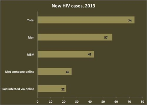
Posted by Acubiz | BlogOf the 74 new HIV cases in Rhode Island in 2013, three in 10 occurred among men who told researchers they believed they were infected by a man met online.
Credit: Brown University
More than 60 percent of Rhode Island men who have sex with men (MSM) diagnosed with HIV in 2013 reported meeting sexual partners online in the preceding year, according to a study published in the journal Public Health Reports.
Study authors at Brown University, The Miriam Hospital, and the Rhode Island Department of Health said companies that produce hookup websites and apps should partner with public health groups, to share public health messages about the risks of sexual encounters arranged online. For instance, sites and apps could provide affordable advertising access to help prevent infection in communities that are most impacted by HIV.
In 2013, 74 Ocean State residents were newly diagnosed with HIV. Three in five were gay, bisexual, or other MSM, and of those 43 people, 22 told researchers they believe a man they met online gave them the virus, according to the study published online in the journal Public Health Reports. The research team interviewed 70 of the state’s 74 newly diagnosed people for the study.
“This is a statewide study that included nearly all individuals newly diagnosed with HIV across an entire state,” said Amy Nunn, associate professor of Public Health and Medicine at Brown University and director of the Rhode Island Public Health Institute. “This is one of the first studies to document how common Internet site use is among people newly diagnosed with HIV and highlights important opportunities to partner with hookup sites to advance public health.”
Five sites and apps, some of which are also used by women, were the most popular: Grindr, Manhunt, Scruff, Adam4Adam and Craigslist. Study lead author Dr. Philip Chan, assistant professor of medicine in the Alpert Medical School and director of at the STD Clinic at The Miriam Hospital, said the widely used sites are part of the lifestyle and culture among many gay and bisexual men and can lead to lasting relationships, not just health risks. The goal of the research, therefore, is not to stigmatize sex or men who use the sites, he and Nunn said, but to instead to inspire partnerships with companies to include more information that could slow the spread of HIV.
“Prevention messaging is a vital tool in our work to prevent new HIV transmissions in Rhode Island,” said study co-author Dr. Nicole Alexander-Scott, director of the Rhode Island Department of Health. “A study like this is an urgent call to action for greater collaboration around education to address the health needs of men who have sex with men. The rate of new HIV diagnoses among men who have sex with men represents an unacceptable health disparity that absolutely must be addressed.”
Seeking public health partnership
“Across the U.S. we are seeing MSM as the number one risk group for HIV infection,” Chan added. “On these online hookup sites, many young MSM are meeting sex partners. It’s really an under recognized and under utilized approach we should be using to reach out to and engage this group.”
To date, public health officials have struggled to sustain informational campaigns on sites and apps that charge for advertising, either because they have no discounts for non-profits or don’t discount enough, Chan said. Craigslist and Scruff ads are free, the authors said, but staff at small non-profit or government agencies face logistical challenges in messaging in these venues, such as having to continually repost ads.
“One of the challenges this study highlights is that it’s prohibitively expensive for many organizations who focus on public health promotion to buy ads on these apps and websites,” Nunn said. “Reducing disease transmission should be part of these organizations’ corporate social responsibility programs.”
The researchers document recent advertising costs in their study, which can quickly run into the thousands of dollars.
“We would like to see more of these companies stepping up to the plate to work with public health departments,” Chan said.
The urgency has not abated since 2013, Chan said. In 2014, the study notes, HIV infections in Rhode Island grew by 97 new diagnoses, again mostly among MSM.
Many of the individuals newly diagnosed in Rhode Island were diagnosed late in the course of their infection, the study showed. Nunn said this suggests that they may have been living HIV for a long time, and potentially unknowingly transmitting HIV to other people, including partners they met online. These findings highlight opportunities to disrupt HIV transmission, she said, by partnering with websites to deliver prevention messaging services that promote routine HIV testing, treatment, and uptake of pre-exposure prophylaxis (PrEP), a once a day pill that can dramatically reduce HIV acquisition risks for HIV negative individuals.
Electron microscopy captures snapshot of structure coronaviruses use to enter cells
Posted by Acubiz | BlogAtomic model suggests vaccine strategies against deadly pandemic viruses such as SARS-CoV and MERS-CoV
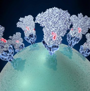
Coronaviruses — the agents behind outbreaks of new kinds of pneumonia — employ molecular tactics to infect cells.
Credit: Veesler Lab/University of Washington
High-resolution cryo-electron microscopy and supercomputing have now made it possible to analyze in detail the infection mechanisms of coronaviruses. These viruses are notorious for attacking the respiratory tract of humans and animals.
A research team that included scientists from the University of Washington (UW), the Pasteur Institute and the University of Utrecht has obtained an atomic model of a coronavirus spike protein that promotes entry into cells. Analysis of the model is providing ideas for specific vaccine strategies. The study results are outlined in a recent UW Medicine-led study published inNature. David Veesler, UW assistant professor of biochemistry, headed the project.
These viruses, with their crowns of spikes, are responsible for almost a third of mild, cold-like symptoms and atypical pneumonia worldwide, Veesler explained. But deadly forms of coronaviruses emerged in the form of SARS-CoV (severe acute respiratory syndrome coronavirus) in 2002 and of MERS-CoV (Middle East respiratory syndrome coronavirus) in 2012 with fatality rates between 10 percent to 37 percent.
These outbreaks of deadly pneumonia showed that coronaviruses can transmit from various animals to people. Currently, only six coronaviruses are known to infect people, but many coronaviruses naturally infect animals. The recent deadly outbreaks resulted from coronaviruses overcoming the species barrier. This suggests that other new, emerging coronavirus with pandemic potential are likely to emerge. There are no approved vaccines or antiviral treatments against SARS-CoV or MERS-CoV.
The ability of coronaviruses to attach to and enter specific cells is mediated by a transmembrane spike glycoprotein. It forms trimers decorating the virus surface. Trimers are structures assembled from three identical protein units. The structure the researchers studied is in charge of binding to and fusing with the membrane of a living cell. The spike determines what kinds of animals and what types of cells in their bodies each coronavirus can infect.
Using state of the art, single particle cryo-electron microscopy and supercomputing analysis, Veesler and his colleagues revealed the architecture of a mouse coronavirus spike glycoprotein trimer. They uncovered an unprecedented level of detail. The resolution is 4 angstroms, a unit of measurement that expresses the size of atoms and the distances between them and that is equivalent to one-tenth of a nanometer.
“The structure is maintained in its pre-fusion state, and then undergoes major rearrangements to trigger fusion of the viral and host membranes and initiate infection,” Veesler explained.
The coronavirus fusion machinery is reminiscent of the fusion proteins found in another family of viruses, the paramyxoviruses, which include respiratory syncytial virus (the leading cause of infant hospitalizations and wheezing in children) as well as the viruses that cause measles and mumps. This resemblance implies that the coronavirus and paramyxovirus fusion proteins could employ similar mechanisms to promote viral entry and share a common evolutionary origin.
The researchers also compared crystal structures of parts of the spike protein in mouse and human coronaviruses. Their findings provide clues as to how the molecular structure of these protein domains might influence which specific animal species the virus is able to infect.
The researchers also analyzed the structure for possible targets for vaccine design and anti-viral therapies. They observed that the outer edge of the coronavirus spike trimer has a fusion peptide — a chain of amino acids — that is involved in viral entry into host cells. The easy accessibility of this peptide, and its expected similarity among a number of coronaviruses, suggests possible vaccine strategies to neutralize a variety of these viruses.
“Our studies revealed a weakness in this family of viruses that may be an ideal target for neutralizing coronaviruses,” Veesler said.
There may be a way, the researchers noted, to elicit broadly neutralizing antibodies recognizing this peripheral peptide. Neutralizing antibodies protect against infections by stopping a mechanism in a pathogen. Broadly neutralizing antibodies would be effective against several strains of pathogen, in this case coronaviruses. The physical structure of the fusion peptide inspires ideas for the design of proteins that would disable it.
“Small molecules or protein scaffolds might eventually be designed to bind to this site,” Veesler said, “to hinder insertion of the fusion peptide into the host cell membrane and to prevent it from undergoing changes conducive to fusion with the host cell. We hope that this might be the case, but much more work needs to be done to see if it is possible.”
The coronavirus spike protein structure described in this Letter to Nature is expected to resemble other coronavirus spike proteins.
“Therefore, the structure we analyzed in the mouse coronavirus is likely to be representative of the architecture of other coronavirus spike proteins such as those of MERS-CoV and SARS-CoV,” the researchers observed.
The researchers summed up their paper, “Our results now provide a framework to understand coronavirus entry and suggest ways for preventing or treating future coronavirus outbreaks.”
“Such strategies,” Veesler said, “would be applicable to several existing coronaviruses and to emerging future strains of coronavirus that conserve this same structure for entering cells.”
How plants protect photosynthesis from oxygen
Posted by Acubiz | BlogDuring the daytime, plants convert the Sun’s energy into sugars using photosynthesis, a complex, multi-stage biochemical process. New work from a team including Carnegie’s Mark Heinnickel, Wenqiang Yang, and Arthur Grossman identified a protein needed for assembling the photosynthetic apparatus that may help us understand the history of photosynthesis back in the early days of life on Earth, a time when oxygen was not abundant in the atmosphere. Their work is published by Proceedings of the National Academy of Sciences.
Photosynthesis takes place in stages. In the ‘first stage’ light is absorbed and used to produce energy molecules, with oxygen as a byproduct. These energy molecules are then used to power the ‘second stage’ of photosynthesis, in which carbon dioxide from the air is fixed into carbon-based sugars, such as glucose and sucrose.
Working with the green alga Chlamydomonas, the research team–which also included graduate students Rick Kim and Tyler Wittkopp of Stanford University, Karim Walters of Pennsylvania State University, and visiting professor Stephen Herbert of the University of Wyoming — focused on a protein, CGL71. It was already known to be involved in assembling the array of proteins that make up the part of the photosynthetic apparatus involved in the first stage of photosynthesis — the one that turns sunlight into the energy molecules that power the second stage and that also has an oxygen byproduct. But little about CGL71’s role in this assembly process was understood until now.
The team was able to figure out that at least one aspect of CGL71’s job is to protect the photosynthetic apparatus from oxygen during its assembly. Yes, that’s right, from oxygen. You see, photosynthesis first evolved in bacteria about 3 billion years ago, a time when the Earth’s atmosphere had very little oxygen. Of course, as photosynthetic bacteria became more and more populous on ancient Earth, the atmosphere changed, eventually creating the oxygen-rich air that we breathe today.
Oxygen is a very reactive molecule that can disrupt the iron-and-sulfur-containing clusters of proteins that are crucial to photosynthesis. Like CGL71, these clusters are critical for the first stage of photosynthesis, where they move electrons in order to create the energy molecules. Just as oxygen can rust iron that makes up a horseshoe or frying pan, it can damage the iron-and-sulfur proteins of the photosynthetic apparatus.
So as oxygen accumulated in the Earth’s atmosphere, the photosynthetic mechanism needed protection from its own byproduct, and CGL71 is one component that evolved to keep the photosynthetic apparatus stable under these new conditions.
“When we look at this critical assembly protein, CGL71, it’s as if we are looking back in time to the era when photosynthetic apparatus had to gradually adjust to the changing atmospheric conditions of our planet,” Grossman said.